This project focuses on understanding how neurons functionwithin broader anatomical networks. Brain regions are extensivelyinterconnected, but these connections are not random—they form topographically organizedpathways that facilitate precise communication over long distances. We are leveragingexisting knowledge of anatomy to study multi-regional interactions in unprecedenteddetail. By combining the latest high-density electrodes with innovations toimprove targeting accuracy, we aim to study how brain-wide networks storeinformation about actions and rewards to optimize future behavior.
Behavior in uncertain environments
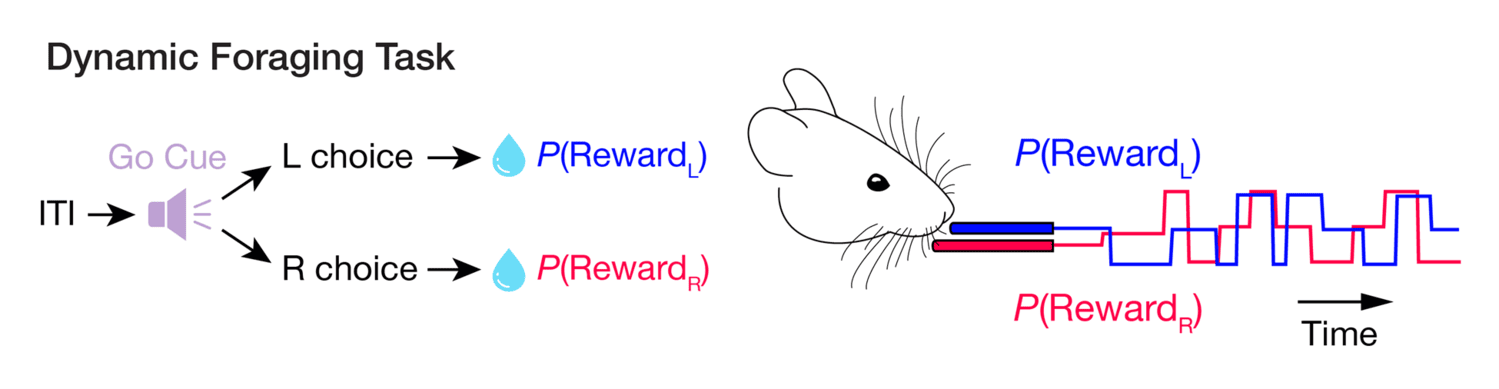
The world around us never stops changing, and our brains must keep track of these changes to ensure we take the right actions at the right time. To study the neural underpinning of this type of flexibility, we train mice to perform tasks where the environmental statistics are constantly in flux, even though the sensory cues remain fixed. This requires subjects to rely on their memory of past actions and rewards to guide future decisions. Previous work has shown that these behaviors depend on widely distributed brain regions, from the frontal cortical areas that track the state of the environment, to the neuromodulatory nuclei that broadcast reward signals, to the loops through basal ganglia that regulate movements. However, very few studies have examined activity across all of these structures in parallel, with single-cell resolution. Our approach focuses on monitoring regions working together in real time, rather than treating them as discrete components that can be observed serially across separate experiments.
Anatomy-guided electrophysiology
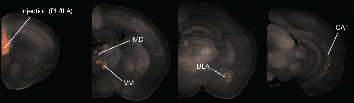
A defining feature of neuroanatomy is its massively parallel structure. Axons extending from every cortical region branch widely, with individual neurons creating links across areas that span the entire brain. Yet nearly everything we know about how neurons behave in living brains stems from experiments that sample only one region at a time. To overcome this limitation, we begin by analyzing data from anterograde, retrograde, and single-cell tracing studies to identify parts of the brain linked by direct projections. Building on prior anatomical surveys, as well as new datasets emerging from the Brain-Wide Anatomy at Cellular Resolution platform, we can build a detailed picture of the inputs and outputs of a cortical region.
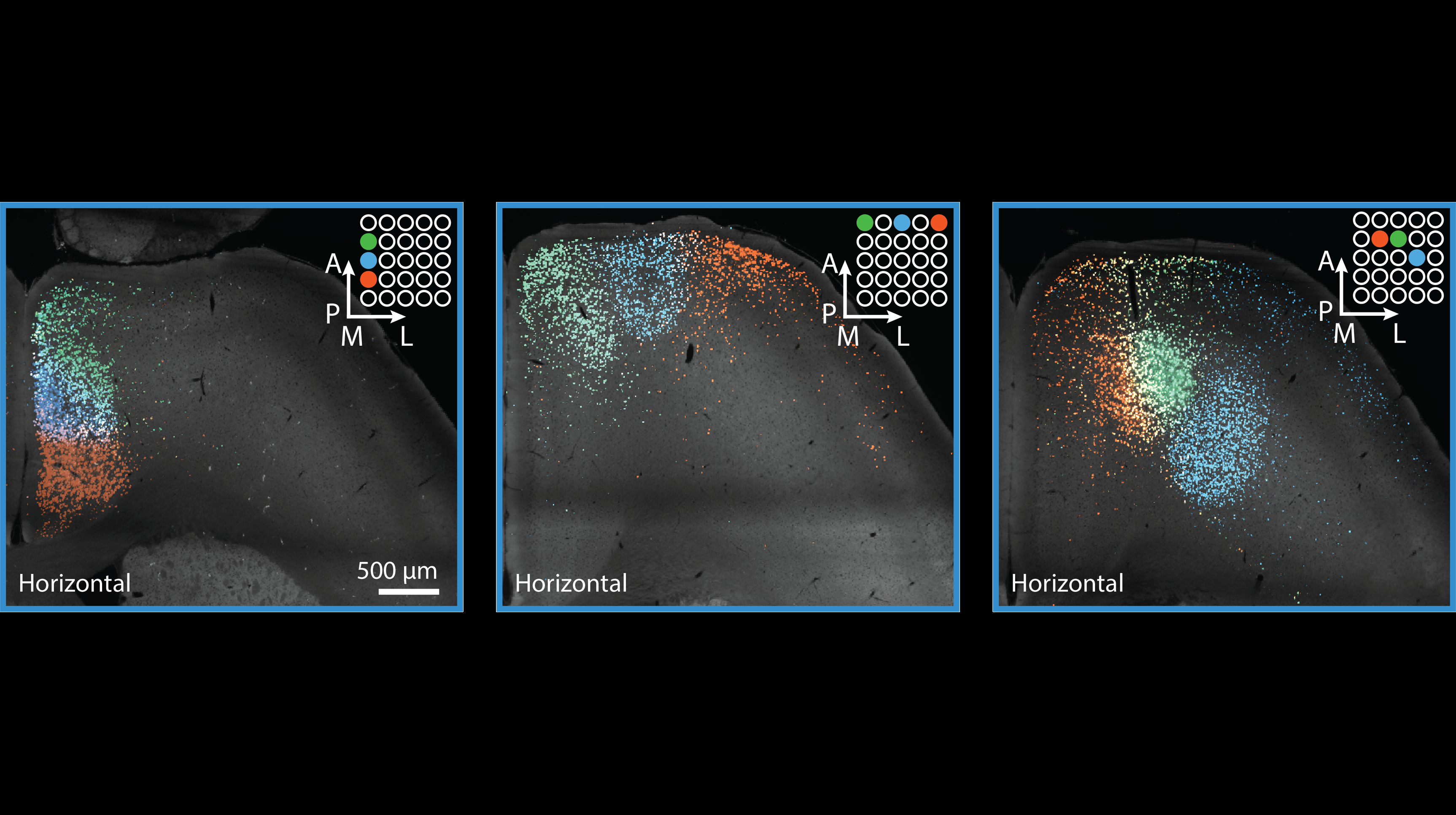
These anatomical maps directly inform our physiological measurements by showing us how to target our electrodes to the most densely interconnected sub-regions of each area. This type of anatomy-guided electrophysiology has historically been challenging due to the variability in electrode placement using standard stereotaxic techniques. However, recent advances made by the Neuropixels Electrophysiology platform—such as the Modular Insertion System and MRI scans from individual mice—have enabled us to target distributed brain regions with remarkable precision and reliability. For example, we can now consistently record from six major inputs to the medial prefrontal cortex in a single experiment. This breakthrough allows us to observe signal propagation across the brain in the context of rich behaviors.
How do multi-regional interactions guide behavior?
Interactive 3D model of a brain-wide electrophysiology experiment, showing 7 Neuropixels probes targeted to the major inputs of prelimbic cortex (PL). The brain volume is extracted from an MRI scan of an individual mouse, allowing us to plan each probe’s trajectory with high precision. Retrogradely labeled cells (from a separate anatomical tracing experiment) are represented as blue dots, highlighting the specific sub-regions that provide the strongest projections to PL.
Our current understanding of brain-wide circuit dynamics has been pieced together from numerous individual studies, leading us to view brain regions as discrete modules rather than as parts of a deeply interconnected system. But abundant anatomical connections ensure that spike-based signals propagate across the brain within milliseconds, making it impossible to trace their origins without precisely targeted recordings from multiple nodes simultaneously. Therefore, we need to move beyond fragmented observations and record simultaneously from major inputs to a given region or from all nodes within multi-regional loops.
Take adaptive decision-making as an example: areas such as prelimbic cortex are known to be essential for behavioral flexibility in changing environments, but the network-level mechanisms of this capability are still largely unstudied. Is the information that guides upcoming actions stored locally within this cortical area, or does it rely on interactions spanning multiple regions? When the cue to act arrives, how does activity cascade through the brain to crystallize into a decision? These questions cannot be adequately answered by studying regions in isolation. Only through anatomy-guided electrophysiology—strategically recording from interconnected sub-regions—can we uncover the principles governing brain-wide communication.
OpenScope Steering Committee
The OpenScope Steering Committee convenes at least biannually to provide crucial direction for the OpenScope project, playing an essential role in ensuring that we effectively serve our broader community.


%20(1).jpg)
.png)