Animals acquire much of their knowledge by freely exploring their environment. Through sustained investigation, they continuously build and update a model of their surroundings: the layout of the space, the objects within it, and how these are organized. For example, rats given free run of a maze will form an accurate map of it even if there is no food at the end; primates learn invariances in the structure of faces just by passive exposure; human infants, after just two minutes of listening to an auditory stream of syllables, will begin to group them into words. Despite its ubiquity, this kind of learning—in which there is no explicit training driven by reward or punishment—has received relatively little attention in systems neuroscience because it is difficult to measure in a nonverbal animal.
This project aims to establish the neural mechanisms that make this form of learning possible. Learning is widely believed to result from changes in synaptic connections between neurons. Current methods for measuring a connectome, the set of synapses in a neuronal network, describe the final state of connections at the animal’s time of death and thus cannot resolve changes related to learning. We are developing a methodology to obtain a living connectome: a time-resolved readout over several weeks of hundreds of synaptic connections in a living brain. The planned research will directly test decades-old hypotheses about the role of synaptic plasticity in learning, such as Hebb’s postulate, and may reveal as yet unimagined mechanisms governing the biological basis of adaptive behavior.
.jpg)
Behavior
We have developed a behavioral paradigm built around the natural drive of mice to investigate their surroundings through their sense of smell. We have found that if we place a port in a mouse’s home cage that delivers neutral odors on demand, mice voluntarily sample from it hundreds of times a day. In this paradigm, the animals have ad libitum access to abundant food and water: there is no task or training, only volitional exploration. We then define a latent rule that determines the exact sequence of odors the animal encounters. By carefully studying spontaneous sampling behavior over weeks, we can infer the rich internal models mice form as they gradually discover this underlying rule. Because the apparatus is simple, automated, and inexpensive, we can run dozens of experiments simultaneously, scaling toward increasingly complex latent structures.
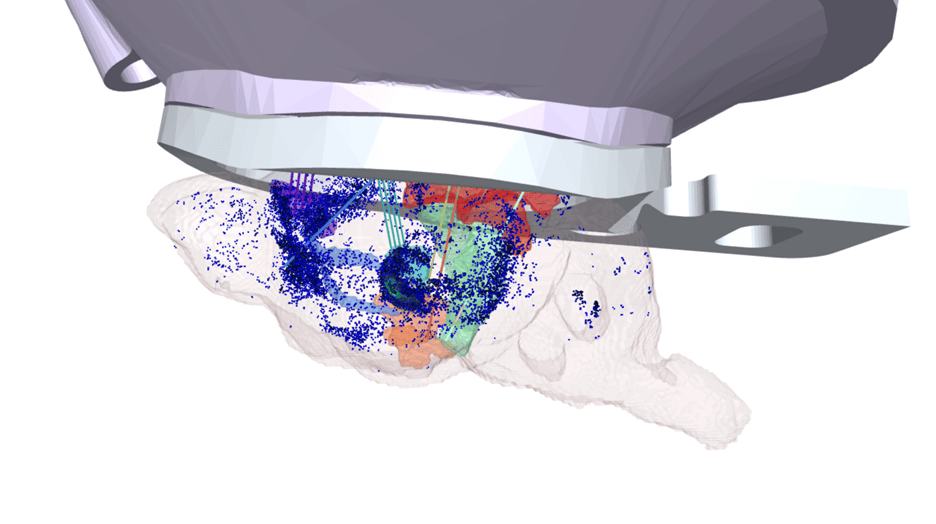
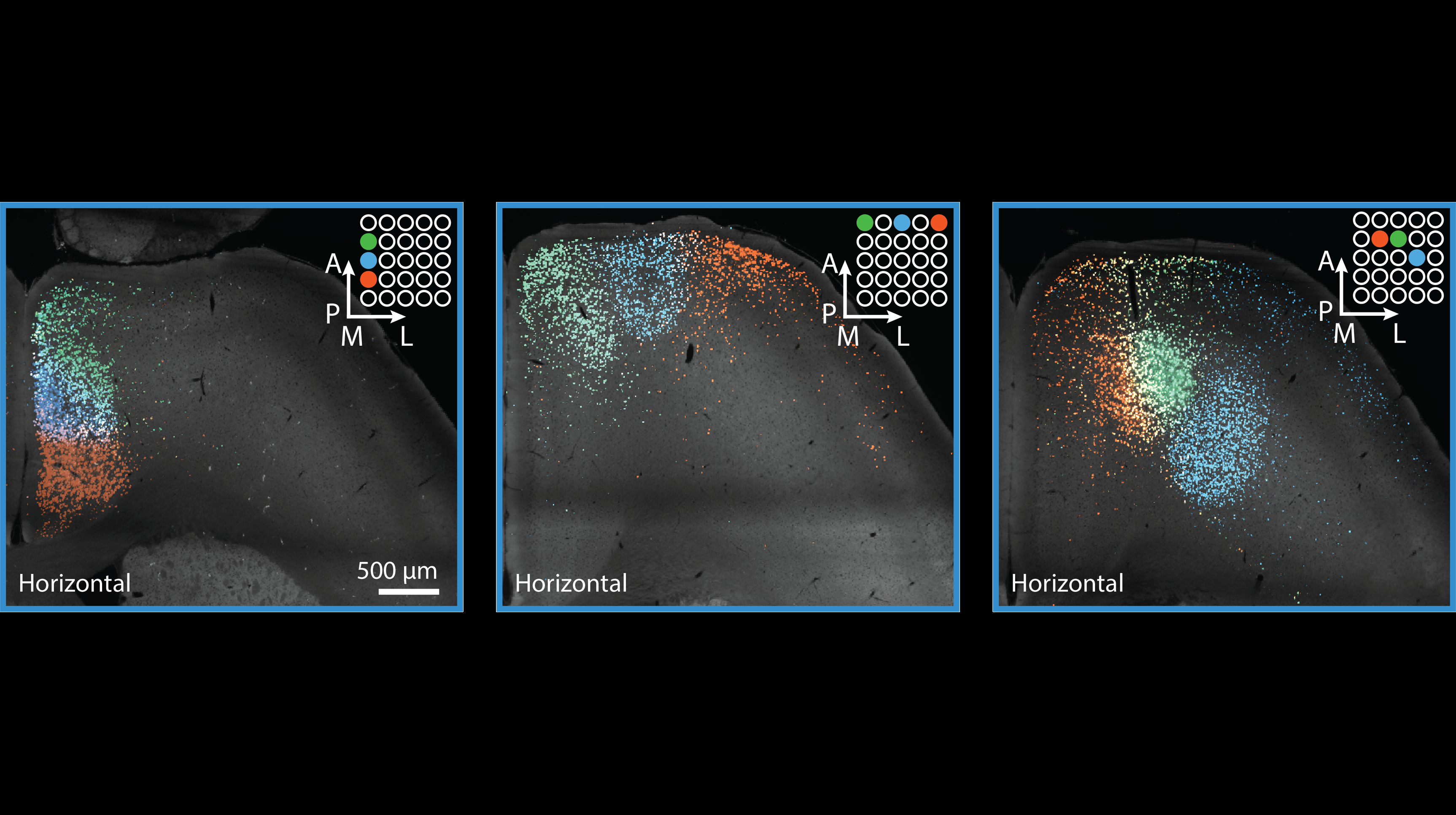
Neural mechanisms
.jpg)
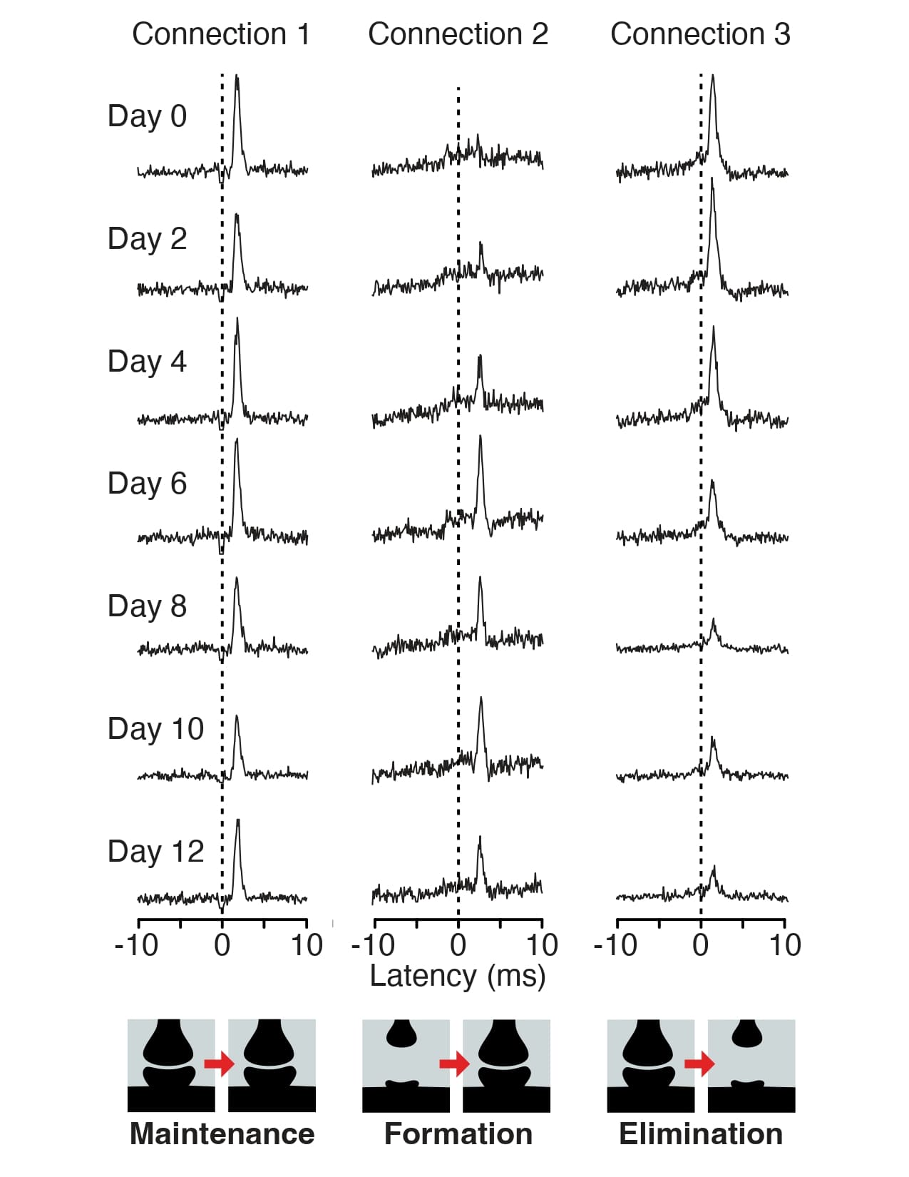
Understanding how the brain supports this learning requires longitudinal, uninterrupted observation of neural circuits over the days and weeks during which the animal freely explores its environment. We aim to measure how a circuit's spiking activity and its synaptic connectivity change together as the animal gradually builds a model of its environment. To this end, we are developing a recording platform to obtain a living connectome: a continuous, multi-week readout of hundreds of synaptic connections alongside the activity of the neurons they connect, measured in a living, learning mouse. This is achieved by combining two capabilities established in our lab: stable, uninterrupted recordings from ~1,000 neurons over ~1,000 hours in freely moving animals; and Dyad, a biophysically constrained machine learning framework that reliably infers monosynaptic connections from extracellular spike trains in vivo. Together, these tools will enable us to directly observe how synaptic connectivity evolves with experience, and to infer the fundamental principles that govern these changes.
OpenScope Steering Committee
The OpenScope Steering Committee convenes at least biannually to provide crucial direction for the OpenScope project, playing an essential role in ensuring that we effectively serve our broader community.


.png)