Some visual neurons have been shown to respond to white noise flicker visual stimuli with high temporal precision, particularly retinal ganglion cells and LGN relay cells. Responses to white noise stimulation are useful for a variety of analyses, including information theoretic measures and generative models of precise spike timing. However the literature was lacking data on responses to white noise in cortical visual areas, or in the visual stream flowing through the superior colliculus.
This experiment used the OpenScope Neuropixels passive viewing protocol, and displayed visual stimuli modulated in time by a short, repeated white noise sequence. The visual stimulus was either a spatially uniform field whose luminance was modulated in time (Full Field Flicker), or a standing sinusoidal grating whose contrast was modulated in time (Static Gratings). Perhaps surprisingly, most cortical visual neurons responded well to full-field flicker white noise. To obtain large populations of neurons in subcortical areas, roughly half of the mice were recorded in a novel electrode configuration.
When white noise visual stimuli are presented repeatedly and the neural responses displayed as spike rasters, the rasters look remarkably like UPC codes or bar codes. The same bar-code-like patterns have been found in neurons recorded in different individual animals, and even neurons in different species. We speculated that these barcodes could be used as identifiers of discrete cell types. The Temporal Barcode Dataset provides "barcodes" for visually responsive neurons throughout the mouse brain, enabling a test of this hypothesis.
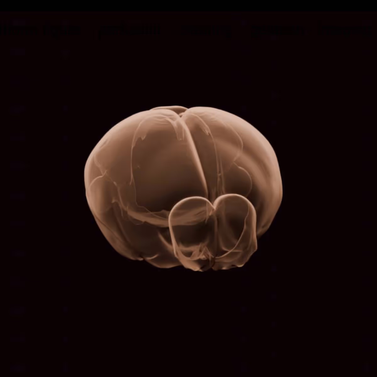
This platform builds on turn-key lightsheet microscopes to image mouse brains at high resolution and throughput. Analysis of the resulting volumetric images, including registration to standard brain coordinates and segmentation and counting of individual neurons, is fully automated.
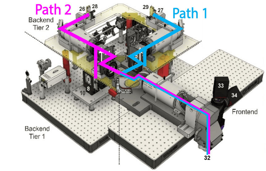
This platform uses a 2-photon microscope SLAP2 that records patterns of synaptic input and output in individual neurons at hundreds to thousands of frames per second in mice performing complex behaviors, using a flexible scan system that combines a digital micromirror device (DMD) with a high-speed scanner.
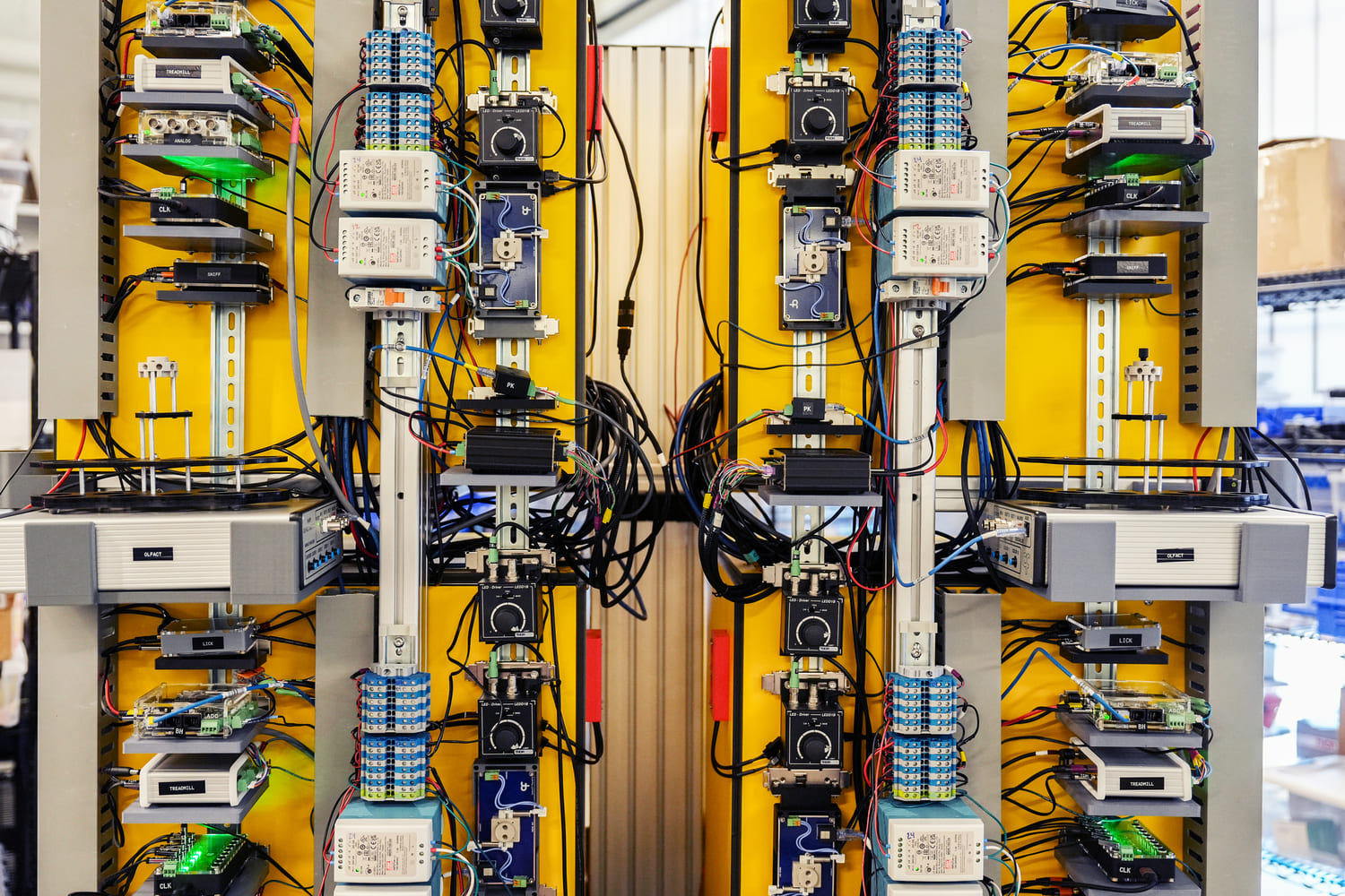
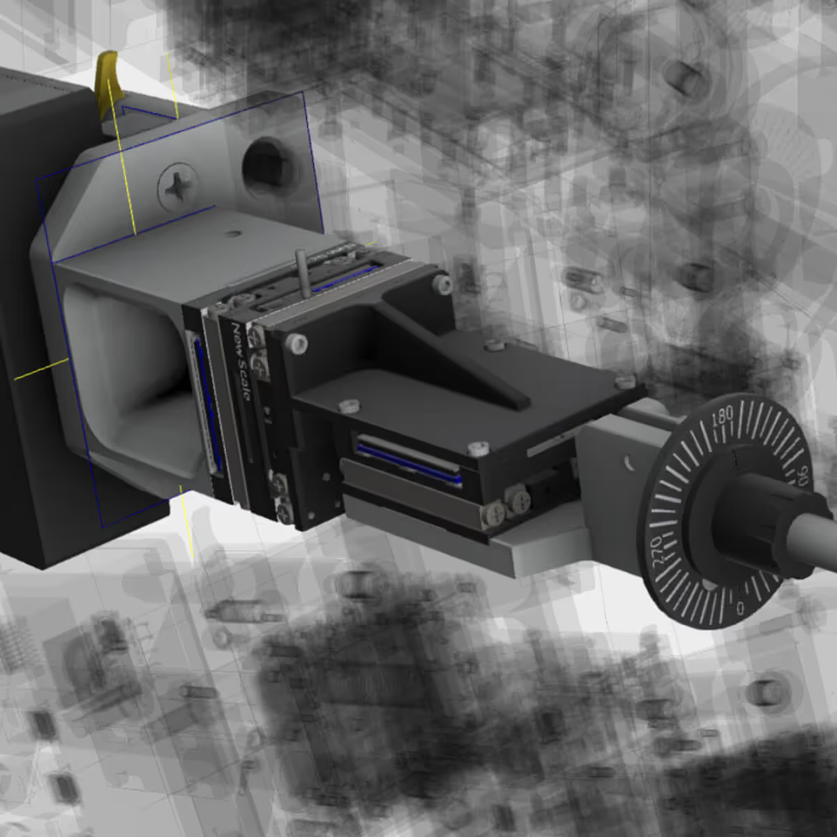
The Scientific Instrumentation and Process Engineering (SIPE) team is a shared engineering resource within the Allen Institute, focused on enabling and scaling cutting-edge bioscience through integrated hardware and software systems.
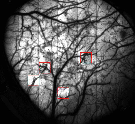
The Population Calcium Imaging platform measures changes in neural dynamics over long time scales as animals learn and behave.
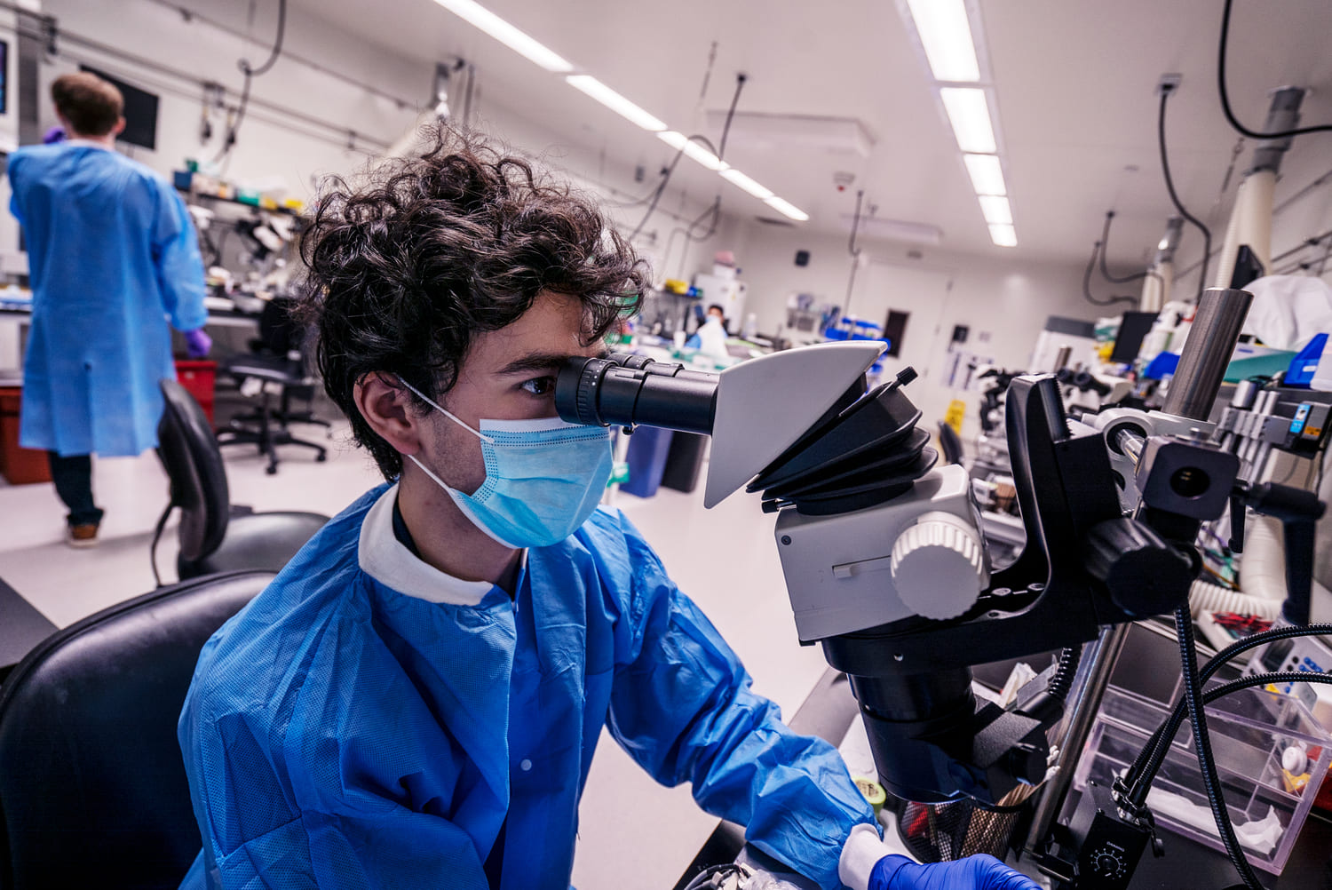
This team performs a variety of surgical procedures, including stereotaxic injections and implanting chronic cranial windows and Neuropixels probes.
This platform implements pioneering technology for highly reproducible, targeted, brain-wide, cell-type-specific electrophysiology to record neural activity from defined neuron types across the brain. Analysis and quality control of the electrophysiology data are fully automated.
This platform combines innovative histology, ExA-SPIM microscopy, image handling, and machine learning to map the morphology and molecular identity of individual neurons across the whole brain at high throughput.
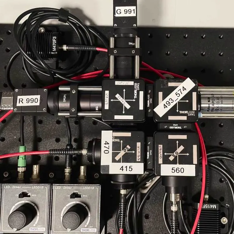
This platform enables optical measurement of neural activity and neurotransmitter release in populations of neurons to study neural circuit dynamics in behaving animals.
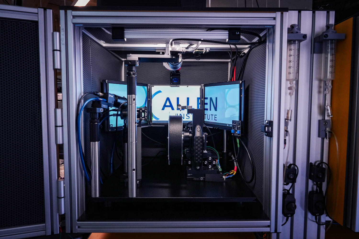
The Behavior platform uses advanced technology to implement a standardized, modular, multi-task virtual reality gymnasium for mice, with the goal to study brain function across different behaviors at scale.