Population Calcium Imaging
Platform
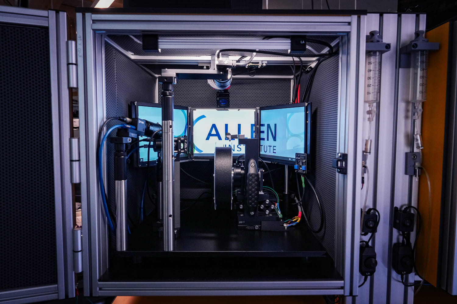
The scientific goals of Neural Dynamics at the Allen Institute rely heavily on an integrated and complementary approach to neurophysiology. While electrophysiological methods afford high-temporal resolution measurements that allow dissection of the neural underpinnings of behavior on the millisecond timescale, calcium imaging provides the opportunity to study changes in neural dynamics over minutes, days, and even weeks as animals learn and behave. By using genetically encoded calcium indicators such as GCaMP, optical physiology provides the unique ability to target and visualize specific genetically defined populations of hundreds to thousands of neurons in each recording session. Being able to visualize cells also enables registration to other image-based data modalities to link in vivo physiology to anatomy or molecular identity of recorded neurons using post-hoc spatial transcriptomics or electron microscopy. Through standardized methods for data acquisition and data processing, we collect diverse datasets for multiple scientific projects in a robust and consistent way. Our pipelines include in-depth quality control of numerous output data streams, detailed tracking of experimental metadata, benchmarked motion correction, segmentation, and event detection algorithms, and packaging of output data into accessible NWB files that we openly share with the scientific community.
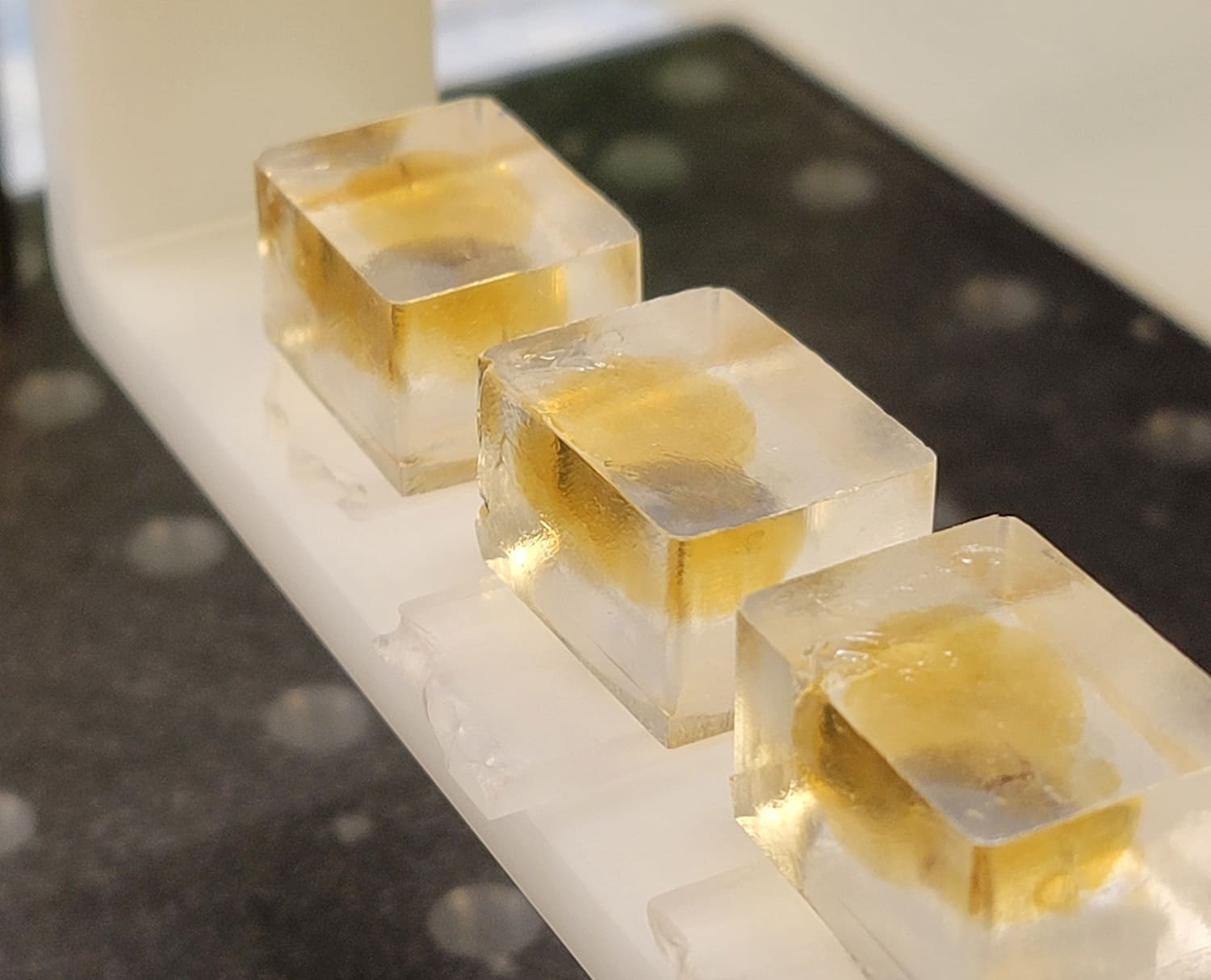
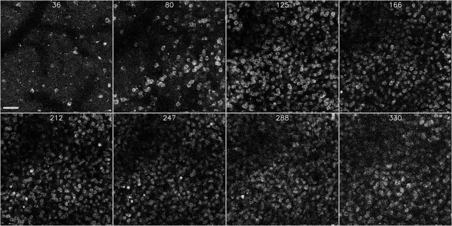
400x400x400µm volume in the primary visual cortex of a mouse expressing GCaMP8s in inhibitory neurons (Slc32a1-IRES-Cre x TIGRE2-jGCaMP8s‐IRES‐tTA2)
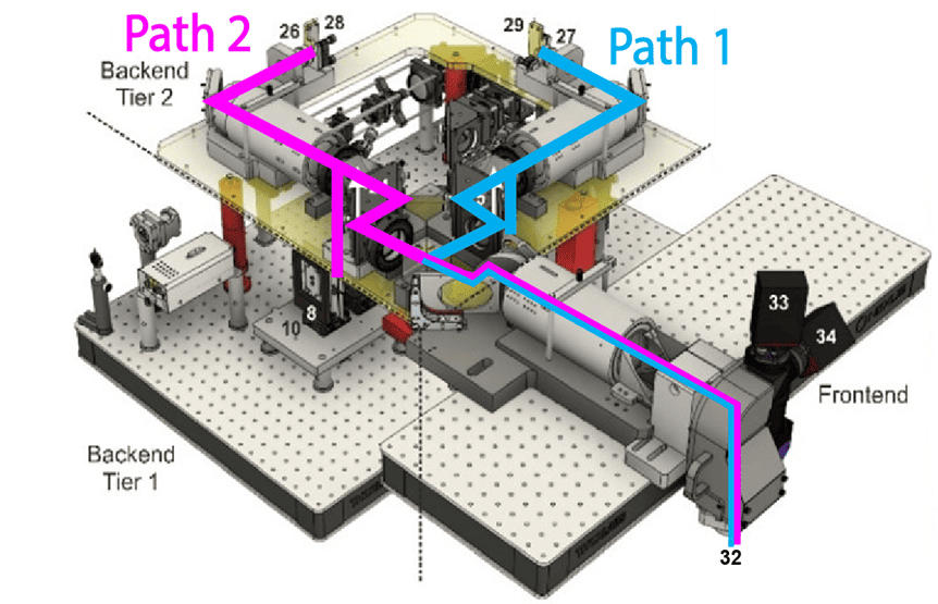
We developed the Multi-plane Mesoscope to address a critical challenge in experimental neuroscience: analyzing spatially distributed neuronal populations with single-cell resolution. Traditional single-plane 2-photon laser scanning microscopy typically does not allow for sampling from distributed neuronal networks across multiple brain regions and cortical depths, limiting both neuronal yield and ability to analyze interareal or intralaminar interactions. Previously, mesoscale imaging systems, such as the 2-photon random access mesoscope (2P-RAM) developed at Janelia Research Campus, have expanded the field of view size, enabling widefield imaging of neuronal activity across multiple cortical areas in a single imaging plane. These systems leverage remote focusing for fast, nearly aberration-free axial scanning.
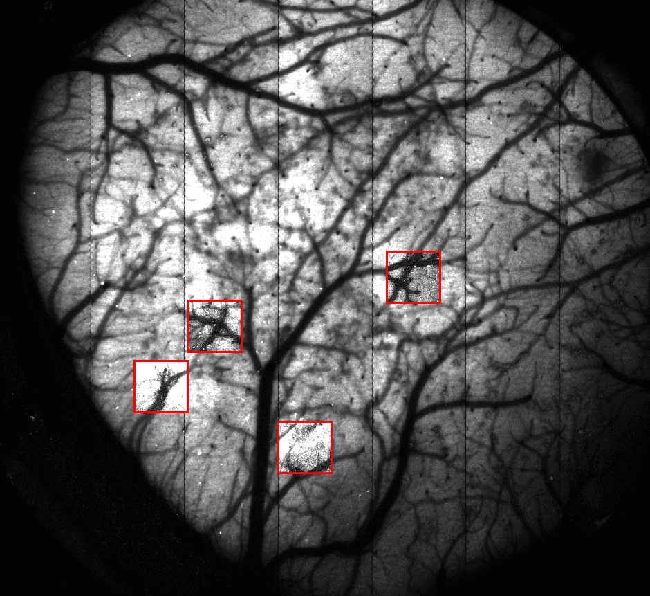
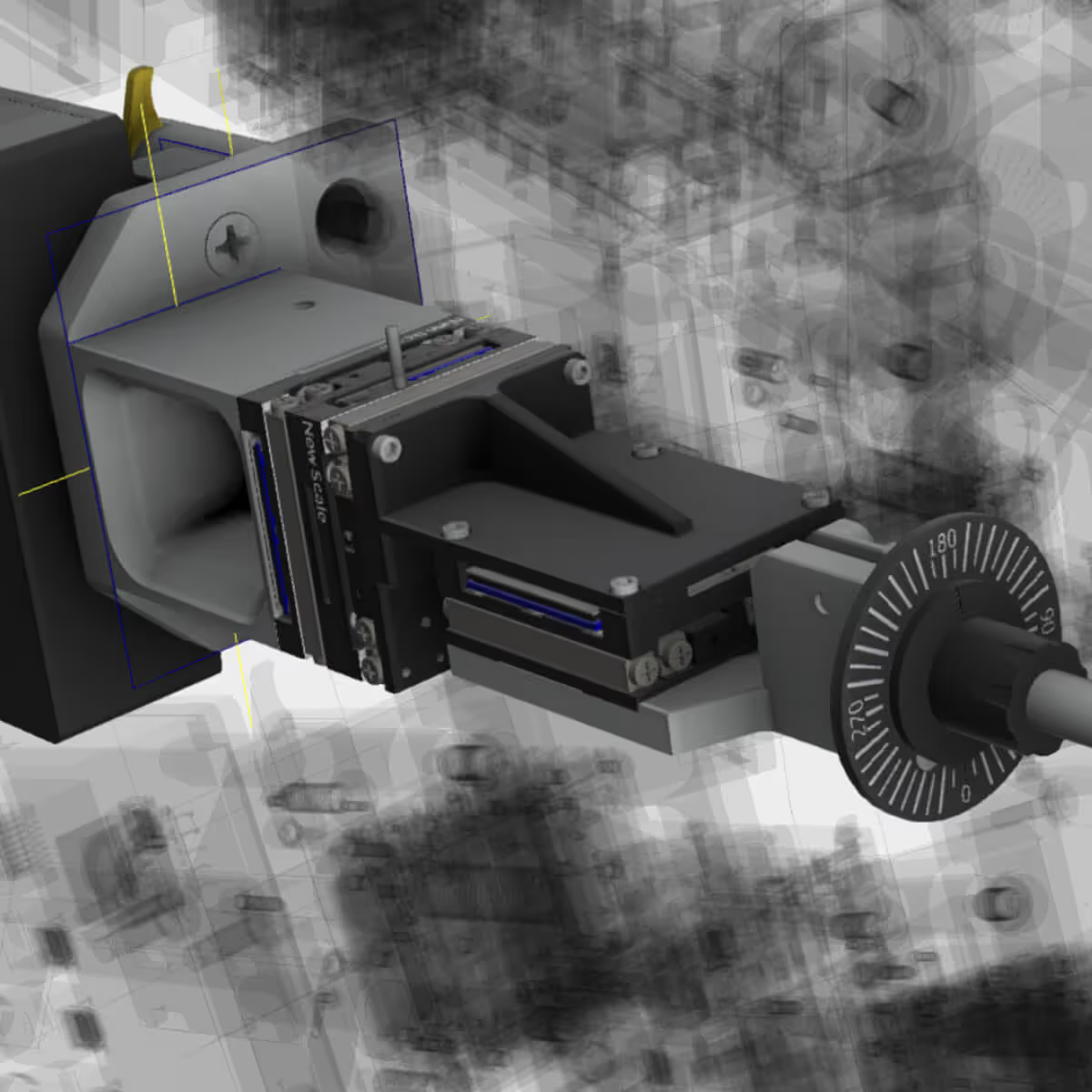
To further expand the throughput of the 2P-RAM system and facilitate study of cortical activity across widespread neuronal networks, the Allen Institute pushed the technology further to develop the Multi-plane Mesoscope, or Multiscope. By combining mesoscopic widefield imaging and spatiotemporal multiplexing, Multiscope effectively doubles imaging output, allowing for simultaneous imaging of up to 8 imaging planes distributed across cortical regions and layers. The Multiscope is commercially available through ThorLabs.
High resolution structural volumes are also acquired to place neurons recorded in individual imaging planes in context of the overall anatomy of the cortical column. These 3D volumes can then be used to align in vivo physiology data to post-hoc histology, spatial transcriptomics, or electron microscopy images acquired from the same tissue sample.
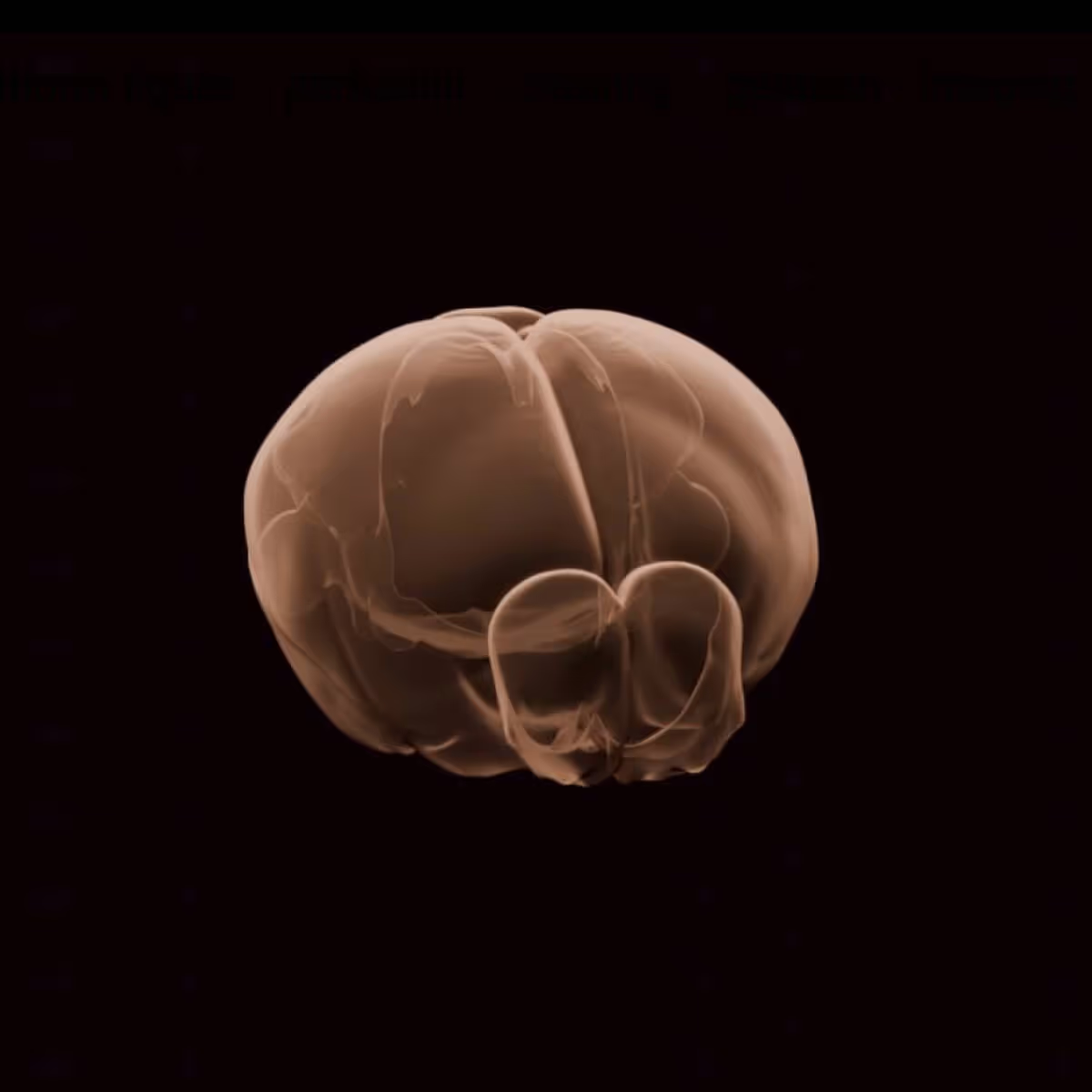
3D rendering of a 400x400x400µm structural volume from the primary visual cortex of a mouse expressing GCaMP8s in all inhibitory neurons (Slc32a1-IRES-Cre x TIGRE2‐ jGCaMP8s‐IRES‐tTA2)
2-photon photostimulation is a powerful technique for precisely perturbing neural activity with cellular resolution that can be combined with population calcium imaging to interrogate circuit dynamics. By leveraging the spatial confinement of 2-photon excitation and genetically expressed opsins for activation or inactivation of neurons, 2P photostimulation enables targeted manipulation of defined neural populations within the intact brain. Simultaneous population calcium imaging enables measurement of the impact of this manipulation on other neurons in the network. We use 2P photostimulation to test causal relationships between neural activity and behavior and measure functional connectivity between neurons.
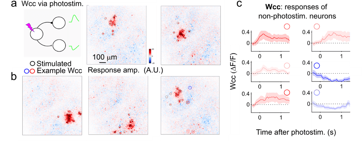
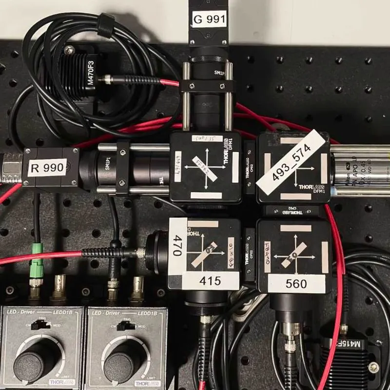
Simultaneous 2-photon imaging and photostimulation requires a dual-laser microscope capable of independently exciting both a fluorescent calcium indicator and an opsin. Our system combines a 920 nm laser for imaging GCaMP-based activity-dependent fluorescence changes with a 1035 nm laser for photostimulating red-shifted opsins (e.g., ChrimsonR and ChRmine). To manipulate large ensembles of neurons, our microscopes employ a spatial light modulator (SLM), which splits the photostimulation beam into multiple beamlets. This configuration enables the simultaneous activation of up to 100 neurons, facilitating systematic perturbations of neural circuits while monitoring the resulting population dynamics.
Movies of calcium fluorescence acquired with our 2-photon microscopes need to be processed to extract the signals of interest from individual neurons. Our optical physiology data processing pipeline takes raw movies coming off the microscopes and performs motion correction, segmentation, neuropil correction, and event deconvolution to extract the corrected, normalized fluorescence traces and event times for each cell in each recording. The pipeline can ingest single-plane or multi-plane experiments and process them according to the specific needs of the data. For instance, multi-plane imaging necessitates the removal of crosstalk between nearby planes, while single-plane imaging does not. Different GCaMP indicators or expression strategies may also have unique needs, such as different segmentation algorithms or neuropil correction considerations. We have designed the pipeline to be highly modular and leverage publicly available tools when possible, including suite2P, CaImAn, OASIS, and ROICat, along with custom solutions as needed. Individual components of the pipeline, such as the segmentation model, can be customized through processing parameters selected by users based on their scientific needs. The pipeline produces several quality metrics to track and quantify algorithm performance to ensure the final output is well validated. The pipeline can be run on the cloud or on local compute clusters. Instructions for using the pipeline with your own data can be found on GitHub.
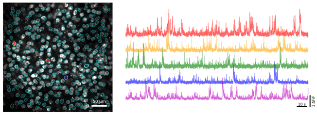
In order to image the activity of specific neural populations with our optical physiology platforms, we use transgenic mouse lines and viral labeling strategies to express calcium indicators in genetically identified populations. GCaMP is a genetically encoded calcium indicator consisting of a green fluorescent protein (GFP) that increases its fluorescence in the presence of calcium ions associated with neuronal activity. We use GCaMP reporter lines that express variants of the GCaMP indicator (e.g., GCaMP6, GCaMP7, GCaMP8) in specific populations of neurons by crossing these reporter lines with various Cre driver lines, or through viral delivery, to target specific genetically defined cell populations depending on the needs of the experiment. The use of transgenic lines in combination with large scale population calcium imaging allows for uniform expression across space (multiple brain areas) and time (multiple weeks of imaging) as mice learn to perform behavioral tasks. Expression of calcium indicators can be combined with other genetic tools to manipulate neural activity, such as opsins for activation or inactivation of neurons through the use of viral expression strategies or a combination of transgenic lines and viruses.
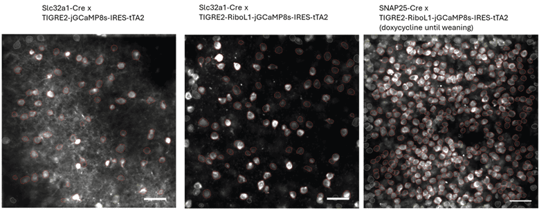
Before using a new GCaMP indicator, transgenic cross, or other expression method, we assess several factors to ensure that the reagents are working as intended for the specific experiments in question, without unintended consequences that could affect data interpretation. These include animal health and behavior, sensor brightness, signal-to-noise ratio, temporal dynamics, and completeness and specificity of GCaMP expression.
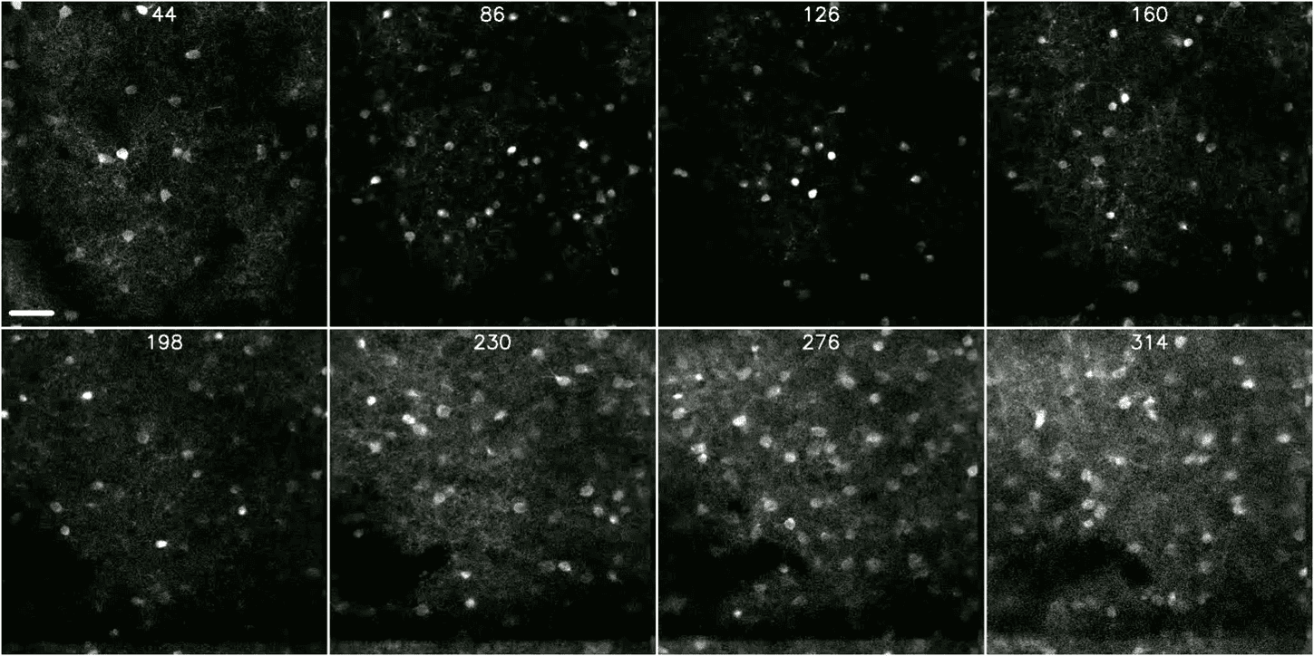
400x400x400µm volume in the primary visual cortex of a mouse expressing Ribo-GCaMP8s in all neurons (SNAP25-Cre x TIGRE2‐RiboL1-jGCaMP8s‐IRES‐tTA2 with doxycycline administered until weaning)
This platform builds on turn-key lightsheet microscopes to image mouse brains at high resolution and throughput. Analysis of the resulting volumetric images, including registration to standard brain coordinates and segmentation and counting of individual neurons, is fully automated.
This platform uses a 2-photon microscope SLAP2 that records patterns of synaptic input and output in individual neurons at hundreds to thousands of frames per second in mice performing complex behaviors, using a flexible scan system that combines a digital micromirror device (DMD) with a high-speed scanner.
The Scientific Instrumentation and Process Engineering (SIPE) team is a shared engineering resource within the Allen Institute, focused on enabling and scaling cutting-edge bioscience through integrated hardware and software systems.
The Population Calcium Imaging platform measures changes in neural dynamics over long time scales as animals learn and behave.
This team performs a variety of surgical procedures, including stereotaxic injections and implanting chronic cranial windows and Neuropixels probes.
This platform implements pioneering technology for highly reproducible, targeted, brain-wide, cell-type-specific electrophysiology to record neural activity from defined neuron types across the brain. Analysis and quality control of the electrophysiology data are fully automated.
This platform combines innovative histology, ExA-SPIM microscopy, image handling, and machine learning to map the morphology and molecular identity of individual neurons across the whole brain at high throughput.
This platform enables optical measurement of neural activity and neurotransmitter release in populations of neurons to study neural circuit dynamics in behaving animals.
The Behavior platform uses advanced technology to implement a standardized, modular, multi-task virtual reality gymnasium for mice, with the goal to study brain function across different behaviors at scale.