OpenScope Vision2Hippocampus project
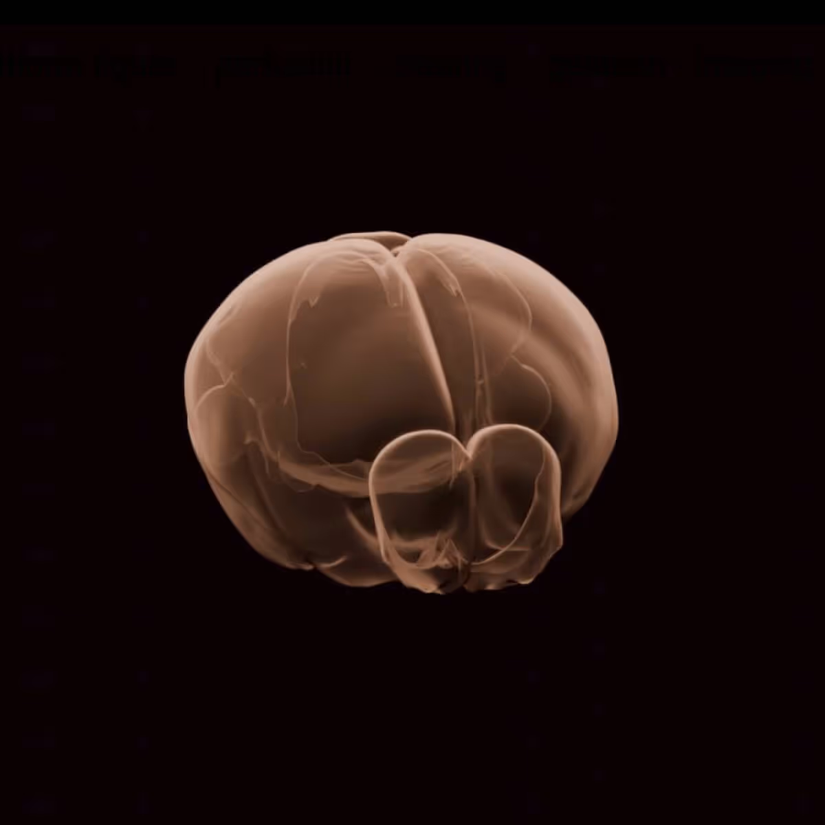
Extensive research shows that visual cortical neurons respond to specific stimuli, e.g. the primary visual cortical neurons respond to bars of light with specific orientation. In contrast, the hippocampal neurons are thought to encode not specific stimuli but instead represent abstract concepts such as space, time and events. How is this abstraction computed in the mouse brain? Specifically, how does the representation of simple visual stimuli evolve from the thalamus, which is a synapse away from the retina, through primary visual cortex, higher order visual areas and all the way to hippocampus, that is farthest removed from the retina?
The current OpenScope project aims to understand how the neural representations of simple and natural stimuli evolve from the LGN through V1, and most hippocampal regions, as well as some of the frontal areas.
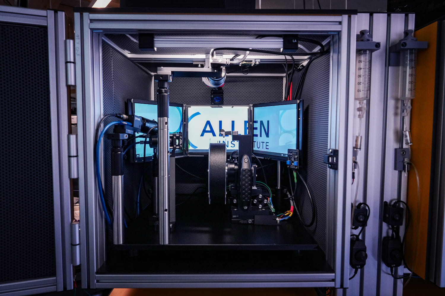
Stimuli presented Two main categories of visual stimuli were presented–
- Simple visual motion, elicited by basic stimuli, like bars of light.
- Complex, potentially ethologically valid visual stimuli, elicited by movies involving eagles (and squirrels).
To parametrize the stimulus properties which might be affecting neural responses, mice were shown variants of the vertical bar of light as follows: A(o) – The bar of light was white, moving on a black background, 15 degrees wide, and moved at a fixed speed, covered the entire width of the screen in 2 seconds. It covered both movement directions consecutively (naso-temporal, then temporo-nasal). A(i) – Similar to A(o), but the bar was now thrice as wide (45o) A(ii) – Similar to A(o), but the bar was thrice as slow (covering the width of the screen in 6 seconds). A(iii) – Similar to A(o), but the contrast was flipped, i.e. a black bar of light on a white background. A(iv) - Similar to A(o), but instead of a simple white bar, the stimulus was striped, and each stripe changed color as the stimulus moved through the width of the screen. This was called “disco” bar of light A(v) – In a subset of mice, A(o) was appended by frames corresponding to the bar of light “vanishing” at either of the edges. Two vanishing protocols were attempted, the bar of light is fully absorbed by the boundary, before reemerging. Another protocol had the bar of light vanish for ~1 second in addition to smoothly being absorbed by the boundary, before reemerging.