Open Tool

Psilocybin collapses visual change detection and drives cortical dynamics toward a state of surprise

Description

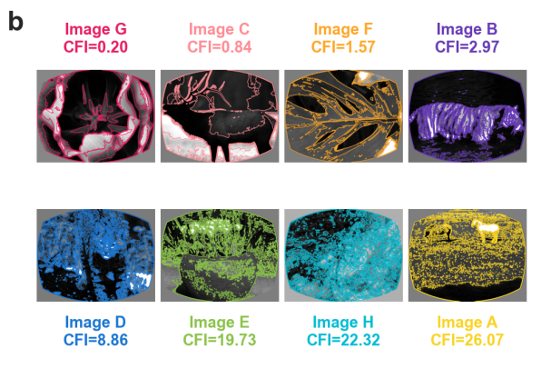
PREPRINT: Psilocybin profoundly alters visual perception, yet the neuronal mechanisms underlying these effects remain unclear. Here we combined large-scale Neuropixels recordings with cell-type-specific optogenetics in head-fixed mice performing a visual change-detection task. Psilocybin severely impaired task performance without overt motor deficits. In cortex, the drug modestly suppressed activity of layer 5 neurons while preserving representations of image identity. By contrast, psilocybin imposed a 4-Hz oscillation on visually evoked activity that preferentially affected neurons encoding image change rather than image identity. Under psilocybin, expected image repetitions aberrantly recruited change-encoding ensembles and shifted cortical population dynamics towards trajectories normally evoked by genuine stimulus changes. These effects were strongest in somatostatin-expressing (SST) interneurons in visual cortex. The strength of this modulation depended on image structure and was greatest for images with clear, continuous contours, which preferentially recruited change-encoding ensembles. These findings demonstrate that psilocybin drives internally generated cortical surprise signals, providing a circuit mechanism for altered perception in the acute psychedelic state.


Projects utilizing this tool

No items found.

Platforms utilizing this tool

No items found.

Platforms accelerate our work

Mesoscale Anatomy

This platform builds on turn-key lightsheet microscopes to image mouse brains at high resolution and throughput. Analysis of the resulting volumetric images, including registration to standard brain coordinates and segmentation and counting of individual neurons, is fully automated.

SLAP2

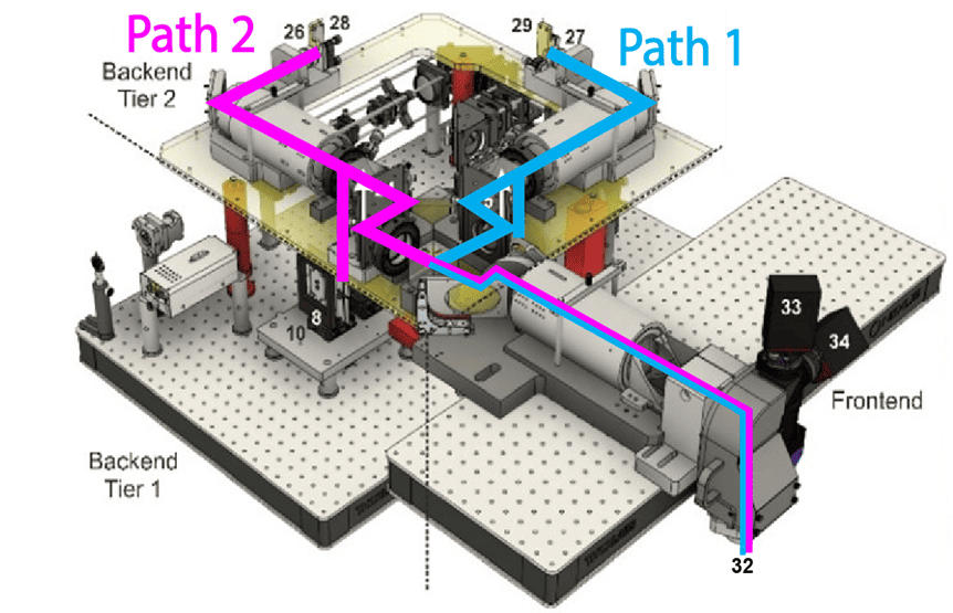
This platform uses a 2-photon microscope SLAP2 that records patterns of synaptic input and output in individual neurons at hundreds to thousands of frames per second in mice performing complex behaviors, using a flexible scan system that combines a digital micromirror device (DMD) with a high-speed scanner.

Scientific Instrumentation & Process Engineering

The Scientific Instrumentation and Process Engineering (SIPE) team is a shared engineering resource within the Allen Institute, focused on enabling and scaling cutting-edge bioscience through integrated hardware and software systems.

Population Calcium Imaging

The Population Calcium Imaging platform measures changes in neural dynamics over long time scales as animals learn and behave.

Surgery

This team performs a variety of surgical procedures, including stereotaxic injections and implanting chronic cranial windows and Neuropixels probes.

Multi-Neuropixels Electrophysiology

This platform implements pioneering technology for highly reproducible, targeted, brain-wide, cell-type-specific electrophysiology to record neural activity from defined neuron types across the brain. Analysis and quality control of the electrophysiology data are fully automated.

Brain-Wide Anatomy at Synaptic Resolution

This platform combines innovative histology, ExA-SPIM microscopy, image handling, and machine learning to map the morphology and molecular identity of individual neurons across the whole brain at high throughput.

Fiber Photometry

This platform enables optical measurement of neural activity and neurotransmitter release in populations of neurons to study neural circuit dynamics in behaving animals.

Behavior

The Behavior platform uses advanced technology to implement a standardized, modular, multi-task virtual reality gymnasium for mice, with the goal to study brain function across different behaviors at scale.